Processing Spectra
Calculating
Lineshape from Data
Angle Resolved XPS Data and Peak Models
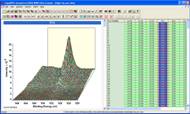
Angle Resolved XPS (ARXPS)
performed using a combination of lens optics and tilting the sample with respect
to the axis for the analyser yield a wealth of data from the same sample. These
data are used to illustrate charge correction via range calibration and further
refined using calibration based on a component peak. Once energy calibrated
these data are processed to form a data set where data bins in each spectrum
are equivalent in a vector sense. These vectors are processed using Principal
Component Analysis and Vector Manipulation to extract peak models for SiO2,
Si2O3 and elemental Si which illustrate the nature of the
native oxide layer on a silicon wafer. Once obtained the peak model is
propagated to spectra over a range of angles.
Start Video Click Here or Click Here for mp4 Mode
Auger Peaks and Determination of Chemical
State
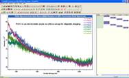
Cu oxide sputtered using an argon
ion gun creates from a nominally Cu 2+ material a range of sub-oxide and
metallic copper within the same sample. A linescan of measurements is performed
by stepping the stage position before acquiring a sequence of spectra to
include Cu 2p, Cu 3p, Cu Auger and valance band spectra. These stage positions
move from a predominantly Cu (0) location to a predominantly Cu 2+ location via
a sequence of stage position increments which trace a path over the edge of the
sputtered zone with the Cu 2+ material. These data are analysed using
calculated lineshapes for Auger spectra corresponding to Cu (0), ion beam
induced Cu sub-oxide and original Cu 2+ material. The analysis involves merging
spectra to form irregular spaced energy spectra, calculation of difference
spectra, PCA as a means of understanding tends within the data set and also PCA
as a noise reduction tool.
Start Video Click Here or Click Here for mp4 Mode
Background
Subtracted Data
Exporting ASCII form of Background
Subtracted Data
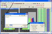
A peak model prepared for an S 2p
doublet complete with loss peaks associated with WS2 2D material is
exported to a spreadsheet program. The exported data appear as columns of ASCII
values corresponding to the background subtracted data together with each
component peak and synthetic envelope resulting from fitting the peak model to
the S 2p spectrum. The example illustrates the process of creating background
subtracted data, copying processed data to a new VAMAS block and extracting an
energy interval as a separate VAMAS block from which the final form of the peak
model is exported through the clipboard.
Start Video Click Here or Click Here for mp4 Mode
Energy
Calibration
Charge Correction and Why Charge Correction is Required in XPS
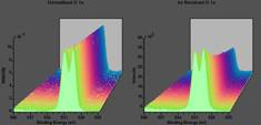
A series of measurements from an
insulating powder is used to illustrate charge compensation during measurements
and how charge correction is performed in CasaXPS. The energy scale is
calibrated by using the binding energy for an O 1s peak to establish the
binding energy scale for five measurements from the same material. An example
of manually calibrating a measurement is illustrated within the video as well
as a demonstration of how energy calibration can be performed automatically for
a set of measurements.
Start Video Click Here or Click Here for mp4 Mode
Binding Energy Calibration and the Influence of Charge Compensation on
Spectra
Binding energy calibration and the
implications of charge compensation for the shapes of photoemission peaks are
examined using PVEE C 1s spectra.
Start Video Click Here or Click Here for mp4 Mode
Range Calibration Option
A set of 10 measurements are
simultaneously calibrated using the C 1s narrow scan spectrum from each
measurement to calibrate other narrow scan spectra from the same measurement.
The measured C 1s peak position is determined using an energy interval over
which the maximum intensity identifies the un-calibrated binding energy for the
as-measured spectra and an offset energy required to place the C 1s peak
maximum at 285 eV is computed and applied en-masse to these 10 independent sets
of spectra within a single VAMAS file.
Start Video Click Here or Click Here for mp4 Mode
Energy Calibration based on Fermi Edge Position
A row of spectra are energy
calibrated based on the position of a Fermi edge. A Step Down background type representing
an approximation to a Fermi edge (in the form of a complementary error
function) is automatically fitted to an edge in the valance band spectra of
aluminium. The position for the Fermi edge, as measured by the intersection of
two straight lines computed from the fitted Step Down background type, is
extracted and used to estimate an energy offset required to locate the edge
position at 0 eV. Spectra corresponding to the same measurement as the valance
band spectrum as calibrated by applying the same offset as is required to
calibrate the Fermi edge.
Start Video Click Here or Click Here for mp4 Mode
Energy Calibration and Data Scaling using a Defined Peak Maximum
Intensity
Energy calibration and data scaling
is performed for a set of measurements where the energy offset and intensity
scale factor for each measurement is calculated from a specified set of
spectra, then applied to all spectra measured under the same conditions. The
VAMAS file consists of a set of VAMAS blocks organised as rows of high
resolution spectra measured under the same conditions. N 1s spectra from each
measurement are used to calculate a binding energy calibration and also a scale
factor. These two adjustments are applied to VAMAS blocks in the same rows of
high resolution spectra collected as part of the same measurement as the N 1s
data.
Start Video Click Here or Click Here for mp4 Mode
Spectrum
Calculator
Summing Spectra with Identical Acquisition Parameters
Data acquired in parallel saved as
separate VAMAS blocks can be combined to form new VAMAS blocks by summing intensities
on an acquisition channel by acquisition channel basis. The example illustrated
in this video demonstrates two methods for summing columns of VAMAS blocks
representing slices within a 2D detector corresponding to two photoemission
peaks with an outcome of two VAMAS blocks for C 1s and Fe 2p spectra. Options
on the Spectrum Processing dialog window Test Data property page are used to
form these two new VAMAS blocks from hundreds of individual VAMAS blocks in the
original data file. Profiling of the signal as a function of detector slice
index is used to select a subset from the total set of VAMAS blocks.
Start Video Click Here or Click Here for mp4 Mode
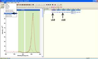
Summing Spectra using the Expression Calculator
A set of spectra are summed using
the shortcut format sum vb<a> :
vb<b> : inc to define the VAMAS blocks for which a sum is required.
Start Video Click Here or Click Here for mp4 Mode
A data set containing nine
acquisition regions split over more than 400 detector slices is used to
illustrate how to energy calibrate four sets of spectra derived from this one
measurement and then sum these energy calibrated spectra to obtain a smaller
FWHM than is obtained by a direct sum based on raw acquisition data channels.
Start Video Click Here or Click Here for mp4 Mode
Normalising Data for Export as ASCII Tables
Exported data in the form of ASCII
tables is performed using the processed data. A normalised form of data can
therefore be exported by first processing VAMAS block data using the Spectrum Processing
Calculator Property Page, where spectral intensities can be adjusted with
respect to a specific energy or at a peak maximum to ensure all data appear
using a common intensity scaling factor.
Start Video Click Here or Click Here for mp4 Mode
Normalising and Scaling Data
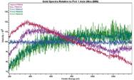
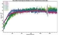
Normalised spectra are scaled using
a common factor to convert a set of spectra with a range of intensities to
VAMAS blocks with a common intensity range. These new VAMAS blocks contain
spectra which permit a display of these data overlaid in a display tile and
also in preparation for further manipulation via the Spectrum Processing
Calculator Property Page. The example used illustrates how to divide a set of
gold spectra by a reference spectrum representing the idealised gold spectrum
from an XPS instrument. The resulting traces provide insight into the
transmission characteristics for the instruments in questions.
Start Video Click Here or Click Here for mp4 Mode
Principal
Component Analysis
Gathering Spectra from Regions defined on VAMAS blocks within a VAMAS
File
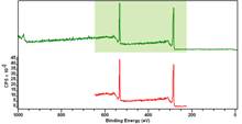
Survey spectra from graphene oxide
(GO) and reduced graphene oxide (rGO) are used to illustrate how to extract
signal between and including O 1s and C 1s photoemission peaks from the
original survey data. After constructing a file containing only the part of these
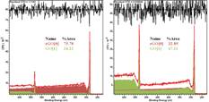
spectra corresponding to O 1s and C 1s, the resulting data are processed using
PCA noise reduction to form a set of spectra with significantly improved signal
to noise characteristics. The results of processing these signal enhanced
spectra are presented as a peak model fitted to the set of survey spectra
allowing a decomposition of the sample into proportions of GO and rGO.
Start Video Click Here or Click Here for mp4 Mode
Image
Calculator
Calculating an Image using Colour Scale Thresholds to Define an
Expression
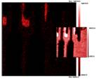
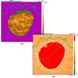
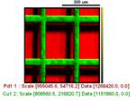
Auger and SEM images are used to
illustrate how contrast within an image can be enhanced and adjusted in terms
of display settings. Once an appropriate pair of threshold values is
established, the image calculation is used to clip intensities above and below
threshold to obtain an image consisting of pixel values within the threshold
range previously prepared for visual inspection of an image.
Start Video Click Here or Click Here for mp4 Mode
Managing
VAMAS Blocks within a VAMAS File
VAMAS Block Order and Re-ordering VAMAS blocks
Data within a VAMAS file are
organised using VAMAS blocks, where each VAMAS block maintains the context for
data within the VAMAS block. When loaded into CasaXPS VAMAS blocks appear as an
array of rectangles with Block Id strings used as labels. The order for these
labelled rectangles is determined by the order for VAMAS blocks within the
VAMAS file and the values assigned to VAMAS block fields. Rows of VAMAS blocks
are ordered by the experimental variable value while columns of VAMAS blocks
are determined by element/transition VAMAS block fields. These VAMAS block
fields can be adjusted within CasaXPS as a means of reordering the arrangement
of VAMAS blocks in the right-hand pane of CasaXPS.
Start Video Click Here or Click Here for mp4 Mode
Linear
Least Squares (LLS)
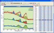
Linear Least Squares applied to Cerium Oxide I
A cerium oxide sample is measured
by splitting the acquisition time over many VAMAS blocks for a set of narrow
scan spectra. Expanding the data set, by recording rapid scans and saving each
rapid narrow scan as a separate VAMAS block, results in 50 spectra per narrow
scan region. Short dwell-times per acquisition bin produces many spectra with
poor signal to noise, but taken as a whole and when summed to create a single
spectrum per photoemission line, the signal-to-noise for these summed spectra
is typical of measurements performed by XPS. PCA and LLS are used to overcome low
counts per bin for these individual spectra leading to hybrid backgrounds
calculated from LLS solutions as well as signal enhanced spectra for each Ce 3d
VAMAS block in the experiment.
Start Video Click Here or Click Here for mp4 Mode
Linear Least Squares applied to Cerium Oxide II
A cerium oxide sample is measured
over an extended acquisition time by repeatedly measuring the same set of Ce 3d
narrow scans using an acquisition time typical for obtaining reasonable signal
to noise in a spectrum. The intension is to induce reduction from Ce 4+ to a
lower oxidation state and by repeatedly measuring the same narrow scan spectrum
have a history of the way these spectra evolve, and to make use of this
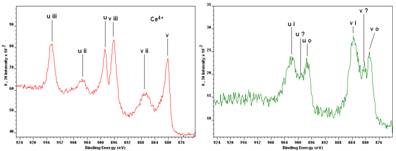
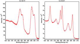
evolution to calculate spectral shapes characteristic of Ce 4+ and Ce 3+
materials. The computed spectra for Ce 4+ and Ce 3+ are compared to spectra
measured from standard materials. These computed Ce 3d spectral forms are then
used within a LLS calculation to demonstrate the suitability of these calculated
spectra for all spectra within the data set.
Start Video Click Here or Click Here for mp4 Mode


